Back Pain Thermal Images
Thermal images can give a strong graphic representation to the underling change before and after ENAR Therapy. Until recently its may have been difficult to ascertain the location of painful areas and related Keypoints. The ENAR is capable of finding and treating these often invisible foci of pain. The thermal image may indicate the before and after effects of treatment.
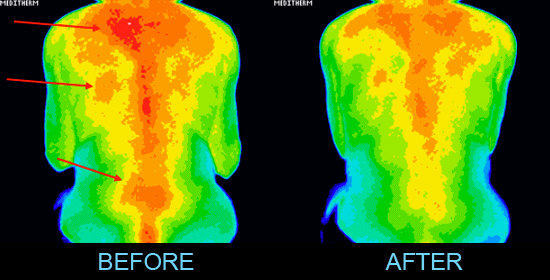
The Case Study patient (above) presented back pain (top to bottom) and left sided posterior chest discomfort. The initial thermal image shows diffuse spine inflammation with discrete focal regions of intense inflammation in the sacrum (bottom arrow), lower thoracic spine and in the musculature medial to the left scapula (top arrow). There is also a region of thermal activity indicating underlying joint dysfunction (second arrow) likely to relate to nerve irritation at the spine. Full spine and paraspinal muscle ENAR Therapy treatment for 30 minutes (single session) demonstrated marked reduction in painful inflammation (hot = red) in all the regions described above which also reflects an improvement in function associated with nerve irritation reversal.